
Figure 3.
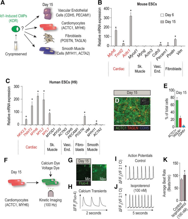
Id1-induced Kdr+ mesoderm is cardiogenic. (A) Schematic depicting the prospective differentiation potential of cryopreserved Id1-induced CMPs to multiple cardiovascular cell types. (B) mRNA expression profiling for the spontaneous differentiation potential of mESCs stably overexpressing Id1 to cardiac (Myh6, Tnnt2, and Actc1), skeletal muscle (Myod1, Myog, and Actn3), vascular endothelial (Cdh5 and Pecam1), and fibroblast (Postn and Vim) markers at days 6 and 15 of differentiation. (C) mRNA expression profiling for the spontaneous differentiation potential of h9 hESCs stably overexpressing Id1 to cardiac (NKX2.5, TNNT2, MYH6, MYL2, and MYL7), skeletal muscle (MYOD1, MYOG, and ACTN3), vascular endothelial (CDH5 and PECAM1), smooth muscle (MYH11 and ACTA2), and fibroblast (POSTN and VIM) markers at days 5 and 15 of differentiation. (D) Representative immunofluorescence image of cardiomyocytes (ACTC1), vascular endothelial cells (CDH5), and fibroblasts (TAGLN) at day 15 of differentiation in h9 hESCs stably overexpressing Id1. Bar, 50 µm. (E) Diagram showing quantification of the percentage of ACTC1+ (cardiomyocytes), TAGLN+ (fibroblasts), and CDH5+ (vascular endothelial cells) at day 15 of differentiation in h9 hESCs stably overexpressing Id1. (F) Schematic of the work flow for the physiological assessment of cardiomyocytes derived from Id1-overexpressing h9 hESCs using the calcium-sensitive (Fluo-4) and voltage-sensitive (VF2.1 Cl) (Miller et al. 2012) dyes. (G) Representative images illustrating the minimum and maximum changes in fluorescence of Fluo-4 in cardiomyocytes derived from Id1-overexpressing h9 hESCs. (H) Representative calcium transient trace of day 15 cardiomyocytes derived from Id1-overexpressing h9 hESCs. (I,J) Representative action potential traces of cardiomyocytes derived from Id1-overexpressing h9 hESCs in control conditions (I) or in response to isoproterenol (J) measured optically with VF2.1 Cl. (K) Beat rate quantification of cardiomyocytes derived from Id1-overexpressing h9 hESCs indicating an increase in beating frequency in response to 100 nM isoproterenol treatment as compared with vehicle and measured with VF2.1 Cl. Quantitative data are presented as means ± SD. All experiments were performed at least in biological quadruplicates. The insets in the top right corners of all immunostaining images shows corresponding DAPI staining.