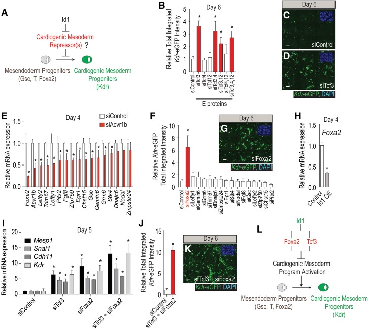
Figure 4.
Id1 promotes cardiogenic mesoderm differentiation by inhibiting Tcf3 and Foxa2. (A) Schematic predicting that Id1 mediates its procardiogenic effect by targeting and inhibiting repressors of cardiogenic mesoderm differentiation. (B) siRNA-mediated functional screen evaluating the role of E proteins (Tcf3, Tcf4, and Tcf12) in repressing cardiogenic mesoderm differentiation. The diagram shows the fluorescence quantification of Kdr-eGFP in response to all seven possible siRNA combinations and siControl. (C,D) Representative immunofluorescence images of Kdr-eGFP at day 6 of differentiation from mESCs transfected at day 3 with siControl (C) and siTcf3 (D). Bar, 50 µm. (E) qRT–PCR validation showing that 17 genes are down-regulated at day 4 in response to siAcvr1b as compared with siControl 24 h after transfection. (F,G) siRNA-mediated functional screen evaluating whether downstream targets of Acvr1b signaling are involved in the repression of cardiogenic mesoderm differentiation. (F) The diagram shows the fluorescence quantification of Kdr-eGFP, where only a siRNA directed against siFoxa2 is able to promote cardiogenic mesoderm differentiation. (G) Representative Kdr-eGFP immunofluorescence images of siFoxa2. Bar, 50 µm. (H) qRT–PCR shows that Foxa2 expression is down-regulated in Id1-overexpressing mESCs as compared with control. (I–K) qRT–PCR for cardiogenic mesoderm markers (Mesp1, Snai1, Cdh11, and Kdr) showing that the cotransfection of siFoxa2 and siTcf3 further enhances cardiogenic mesoderm differentiation as compared with siTcf3 or siFoxa2 alone (shown in I). The diagram shows the fluorescence quantification of Kdr-eGFP (J) and a representative image (K) of the siTcf3 + siFoxa2 condition. Bar, 50 µm. (L) Model showing Id1's repressive role on Tcf3 and Foxa2 activity to promote cardiogenic mesoderm differentiation.

