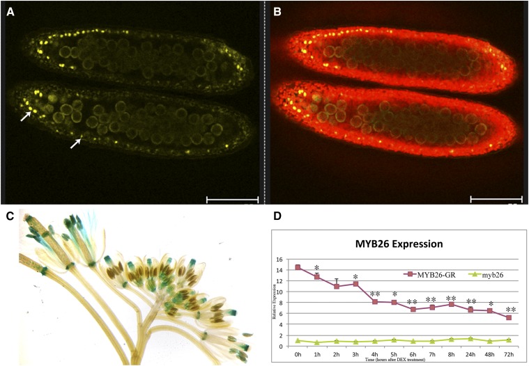
Figure 2.
Localization of MYB26 after DEX-induced expression. A and B, Confocal imaging of expression of the functional MYB26pro:MYB26-YFP fusion protein in anthers; expression is only seen in the nuclei of the anther endothecium cells during pollen mitosis I. A, MYB26-YFP fusion protein localized in endothecium nuclei (arrows; excitation 514 nm). B, Overlay of anther chlorophyll autofluorescence (excitation 488 nm) and MYB26-YFP fusion protein. Scale bar represents 75 µm. C, MYB26Pro:GUS expression is seen in many floral tissues, including nectaries, style, filaments, and anthers. D, Time course of MYB26 expression by qRT-PCR in myb26 mutant buds and myb26 mutant carrying the MYB26pro:MYB26-GR-YFP transgene after DEX treatment. Expression levels of the transgene fluctuated slightly but were reduced 1 h post-DEX treatment and strongly reduced by 4 h post-DEX treatment with all samples being at least P < 0.05 after 3 h compared to 0 h control (t test statistical analysis; *P ≤ 0.05; **P ≤ 0.01).

