Figure 1.
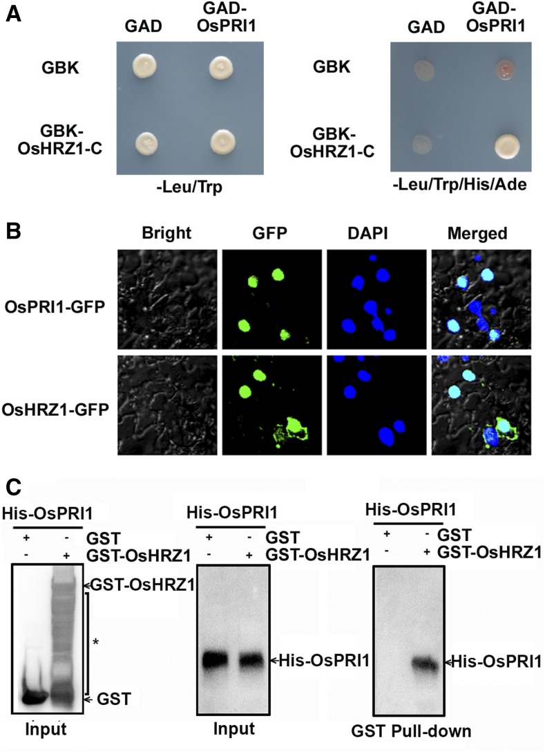
OsPRI1 interacts with OsHRZ1. A, Yeast-two-hybrid assay. Interaction was indicated by the ability of cells to grow on synthetic dropout medium lacking Leu/Trp/His/Ade. The C-terminal truncated OsHRZ1 and full-length OsPRI1 were cloned into pGBKT7 (GBK) and pGADT7 (GAD), respectively. B, Subcellular localization. OsPRI1 and OsHRZ1 were respectively fused to the N-terminal of GFP protein. Fluorescence was observed in nuclear compartments of N. benthamiana leaf epidermal cells. The 4,6-diamidino-2-phenylindole (DAPI) staining indicates the localization of nuclei. Merged images show colocalization of GFP and DAPI signals. C, Pull-down assay. OsHRZ1 was fused with the GST tag, and OsPRI1 was fused with the His tag. Recombinant proteins were expressed in E. coli. Proteins were pulled down by glutathione Sepharose 4B and detected using the anti-His or anti-GST antibody. The asterisk indicates nonspecific signals.

