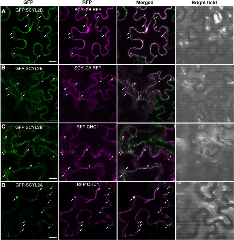
Figure 8.
SCYL2B colocalizes with SCYL2A and CHC1. Tobacco leaf epidermal cells were coinfiltrated with A. tumefaciens strains carrying the following constructs: GFP:SCYL2B and SCYL2B:RFP (A), GFP:SCYL2B and SCYL2A:RFP (B), GFP:SCYL2B and RFP:CHC1 (C), and GFP:SCYL2A and RFP:CHC1 (D). The localization of fluorescent proteins was examined by the detection of GFP and RFP signal by confocal microscopy. Representative images of each sample are shown. White color indicates strong colocalization. Arrows indicate spots showing colocalization. Bars = 10 μm.

