Figure 1.
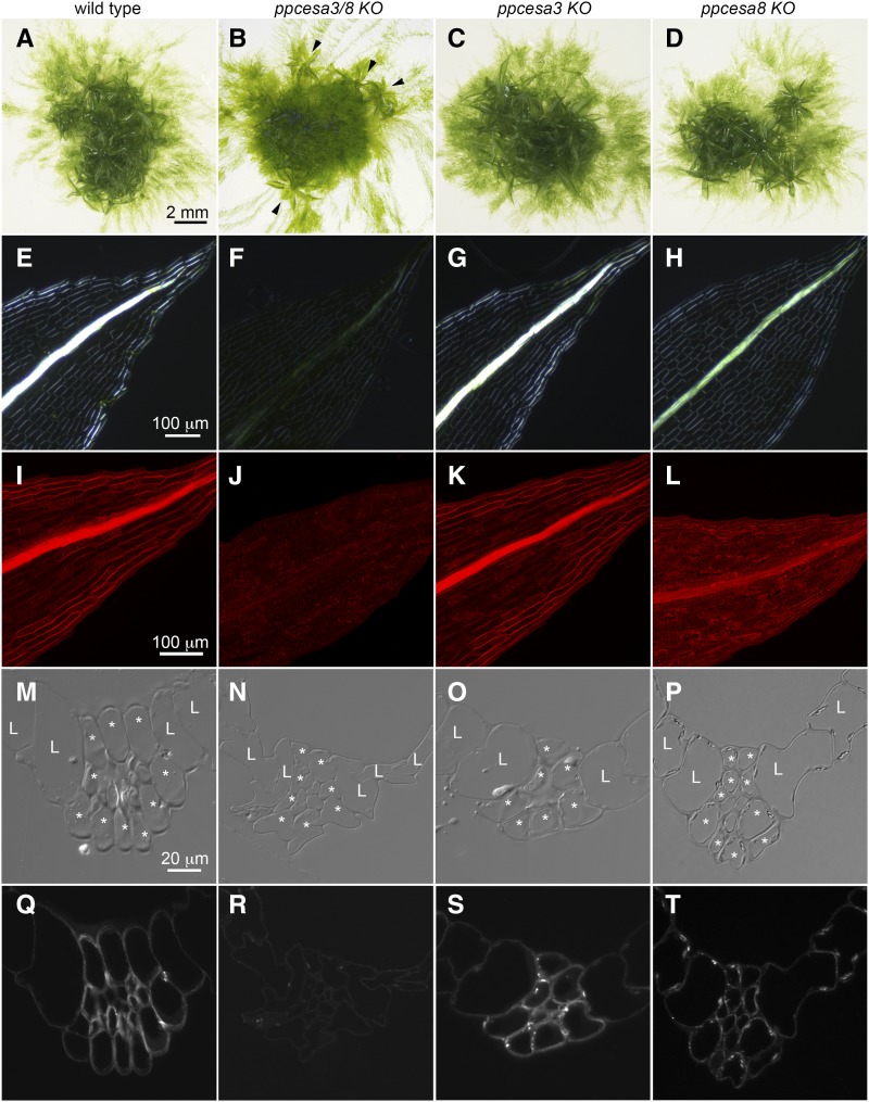
Phenotypes of ppcesa3/8KO, ppcesa3KO, and ppcesa8KO compared with wild type P. patens. A to D, Colony morphology is similar in the wild type, ppcesa3KOs, and ppcesa8KOs; horizontal growth is typical of gametophores produced by ppcesa3/8KO (arrowheads). E to H, Polarized light microscopy of leaves shows that the midribs of the wild type and ppcesa3KO are highly birefringent. The midribs of ppcesa3/8KO leaves have low birefringence, and ppcesa8KO midribs have moderate birefringence. I to L, Fluorescence microscopy of leaves stained with Pontamine Fast Scarlet (S4B) shows strong fluorescence in the midribs of wild type and ppcesa3KO leaves, low fluorescence in the midribs of ppcesa3/8KO leaves, and intermediate fluorescence in the midribs of ppcesa8KO leaves. M to P, Differential interference contrast microscopy of sections through the midribs of maturing leaves (L, lamina cell; *, bundle sheath cell). In the wild type and ppcesa3KO, the walls of bundle sheath cells and the stereid cells they surround show enhanced contrast due to differential refractive index. Q to T, Fluorescence microscopy of the same sections shown in M to P labeled with CBM3a. The bundle sheath and stereid cells of wild type and ppcesa3KO leaves are strongly labeled, whereas labeling is weak in ppcesa3/8KO and intermediate in ppcesa8KO leaves.

