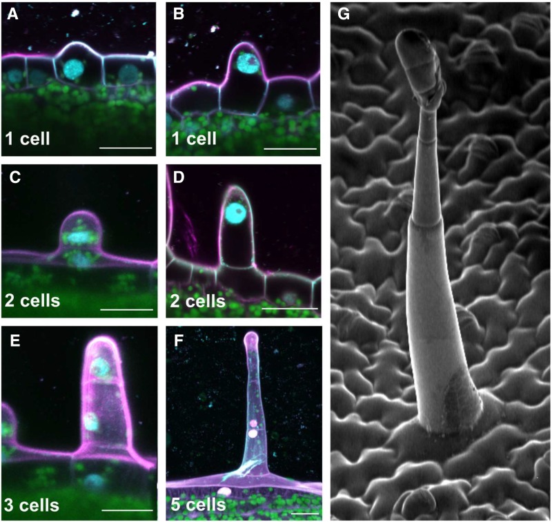
Figure 2.
Glandular trichome initiation and development in N. tabacum. A to F, Confocal microscopy images showing the early steps of glandular trichome development. The number of cells forming the developing glandular trichome is shown at the bottom of each frame. A differentiating protodermal cell enlarges and forms a protuberance (A), the cell nucleus migrates to the tip of the protuberance (B), and cell division takes place (C), forming a structure made of two cells (D). The upper cell protruding from the epidermis then undergoes an asymmetric division, forming one large cell (which will form the multicellular stalk after several rounds of controlled cell division) and one small cell (which will give rise to the multicellular glandular head; E). A developing trichome made of five cells is shown in F. Scale bars, 20 μm. Magenta represents cell wall (propidium iodide staining), cyan represents nuclei (4',6-Diamidine-2'-phenylindole staining), and green represents chloroplasts (chlorophyll a autofluorescence). G, Scanning electron micrograph showing the typical cell architecture of a mature long glandular trichome.

