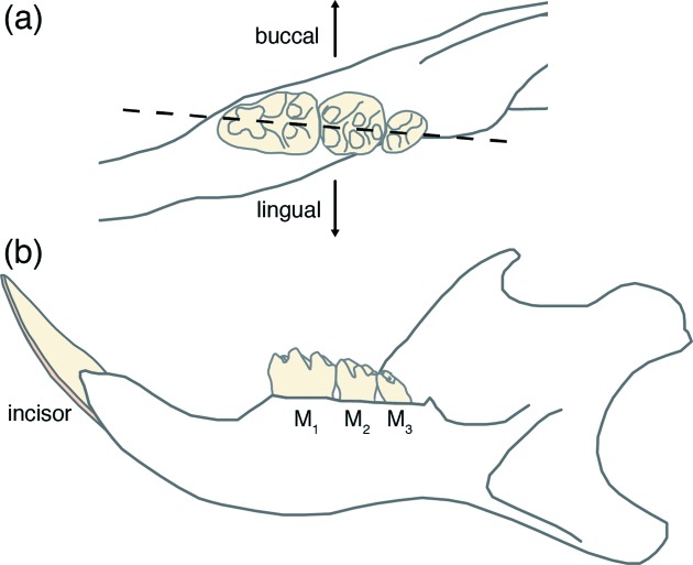
Figure 1.
Schematic drawing of the right mandible of the rat. (a) Enlarged occlusal (top) aspect of the three molars M1, M2 and M3, and the mesio-distal plane (dotted line) along which hemisections were prepared for caries lesion scoring after Keyes (1958 ▸). (b) Lingual (tongue-facing) aspect of the whole mandible.

