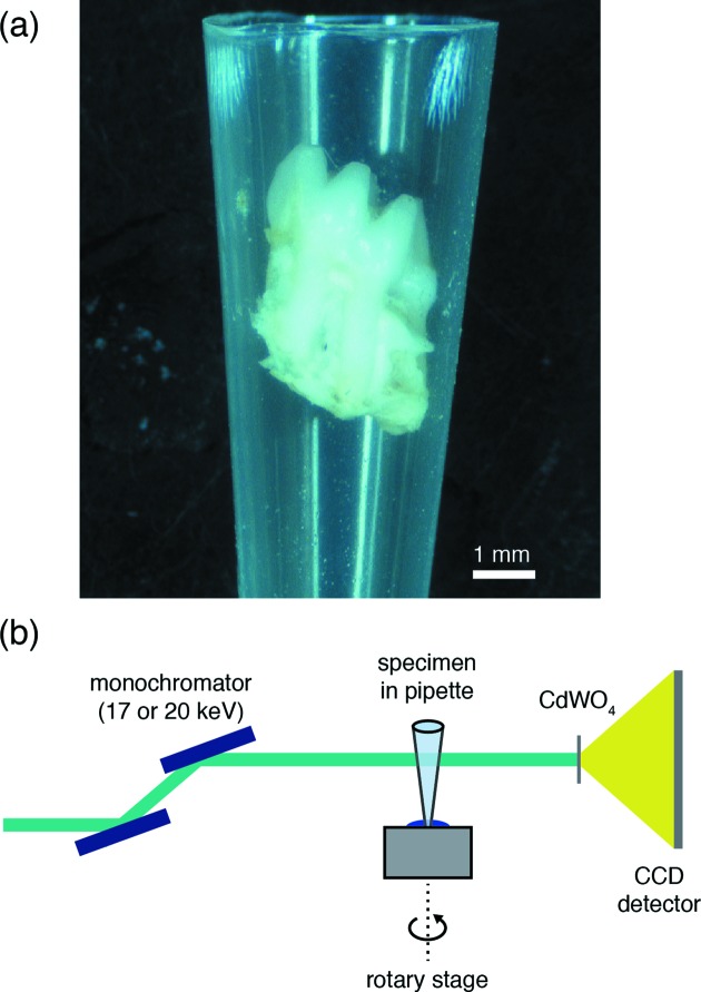
Figure 2.
Sample mounting for synchrotron µCT. (a) The M1 molar wedged inside a modified pipette tip. (b) Schematic drawing of the experimental setup for data collection. A Si(111) double-crystal monochromator selects the beam energy. The specimen in the pipette tip is mounted on the rotation stage using a small piece of modelling clay. A cadmium tungstate (CdWO4) single-crystal scintillator converts X-rays to an optical wavelength, which are then magnified by a 4× objective lens (numerical aperture of 0.2) and collected by CCD detector.

