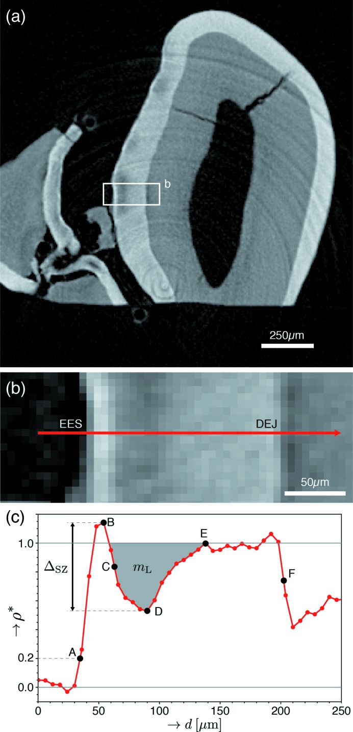
Figure 4.
Definition of parameters used to characterize SZ lesions. (a) Contrast-adjusted slice containing mild lesions within the grooves between tooth cusps (sulcal surfaces). (b) Enlarged image of the boxed region in (a). (c) Plot of the fractional mineral density (ρ*) against distance, taken along the red arrow in (b). Points used for profile quantification were defined as follows: external enamel surface (EES) (A), corresponding to ρ* = 0.2; maximum (B), midpoint (C) and minimum (D) of ρ* within the SZ and lesion; recovery to sound enamel (ρ* = 1) (E); and DEJ (F). The midpoint (C) is defined as the depth at which ρ* equals the average of that at points B and D. These points were selected following the approaches of Groeneveld & Arends (1975 ▸) and Cochrane et al. (2012 ▸) to facilitate direct comparison with human SZs. From these points, depth measurements were calculated for each lesion, including multiple representations for the SZ thickness (AB, AC and AD), total lesion depth (AE) and enamel thickness (AF). Furthermore, the SZ magnitude, ΔSZ, was defined as the difference in ρ* between points B and D. Finally, the integrated demineralization (shaded gray), m L, is defined as the integrated value of 1 − ρ* (normalized mineral lost) within the body of the lesion (where ρ* < 1).

