Figure 3.
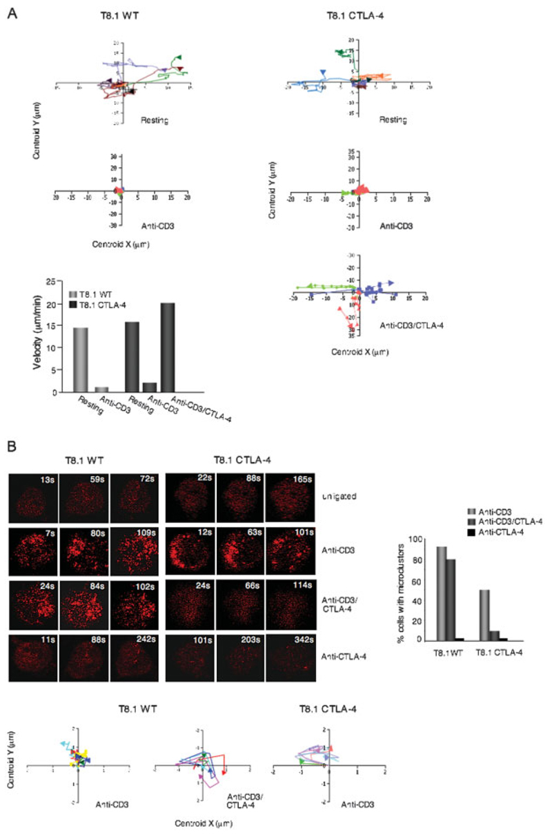
CTLA-4 co-ligation increased cell motility on antibody-coated glass slides while reducing the formation of ZAP70 microclusters. (A) Upper left and right panels: XY tracker charts showing examples of movement of individual microclusters formed in T8.1 WT and T8.1-CTLA-4 cells when left untreated or stimulated with anti-CD3 or anti-CD3/CTLA-4. Lower left panel: T8.1-CTLA-4 cells show increased motility. T8.1 WT and T8.1-CTLA-4 cells transfected with mRFP-ZAP70 were plated in their culture medium on glass slides coated with anti-CD3 or anti-CD3/CTLA-4 and monitored for movement using a Zeiss LSM 510 confocal microscope. Velocity software was used for measuring velocity. (B) Left upper panel: T8.1-CTLA-4 cells show defective ZAP70 cluster formation in response to anti-CD3/CTLA-4 ligation. T8.1 WT and T8.1-CTLA-4 cells transfected with mRFP-ZAP70 were plated in their culture medium on glass culture slides coated with anti-CD3, anti-CD3/CTLA-4 or anti-CTLA-4. Cells were imaged for the formation of ZAP70 microclusters at the interface for a period of time using a Zeiss LSM 510 confocal microscope. Right panel:histogram showing the percentage of cells forming ZAP70 microclusters. Left lower panels: XY tracker charts showing microcluster movement of T8.1 WT cells treated with anti-CD3 or anti-CD3/CTLA-4, and T8.1-CTLA-4 cells treated with anti-CD3. Similar results were obtained from at least two other independent experiments.

