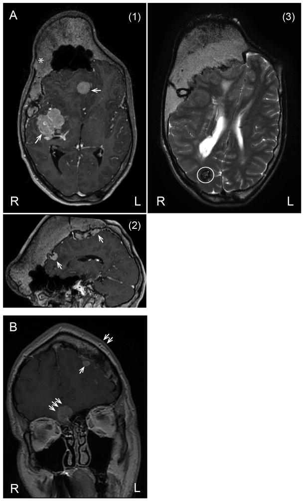
Figure 2.
33 year old with PS, (A) (1) Axial and (2) sagittal views on the MRI scan shows hyperostosis crossing the midline, predominantly involving the right side, meningiomas (thin arrows), and (3) another axial view shows one cavernoma or capillary telangiectasia (circled). 34 year old with PS, (B) MRI scan shows left frontal bone hyperostosis (double arrows) with adjacent meningioma (thin arrow); there is second meningioma (triple arrows) without hyperostosis adjacent to the sphenoid bone.

