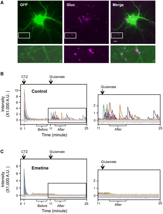
Figure 2. De Novo Arc Translation Is Detected by Gluc.

Substrate (CTZ) was added at 1 min and glutamate was added at 11 min.
(A) Gluc signals after glutamate addition were stacked and overlaid to EGFP signal. Bottom row shows the magnified pictures from the white square in the upper row. The intensity of EGFP signal in the magnified pictures is increased to show the dendritic morphology. Scale bar, 10 μm. (B and C) Discrete spikes were manually selected and individual signal intensities were plotted in different colors. The right is the magnified graph from the box in the left graph. (B) Control. (C) Emetine.
