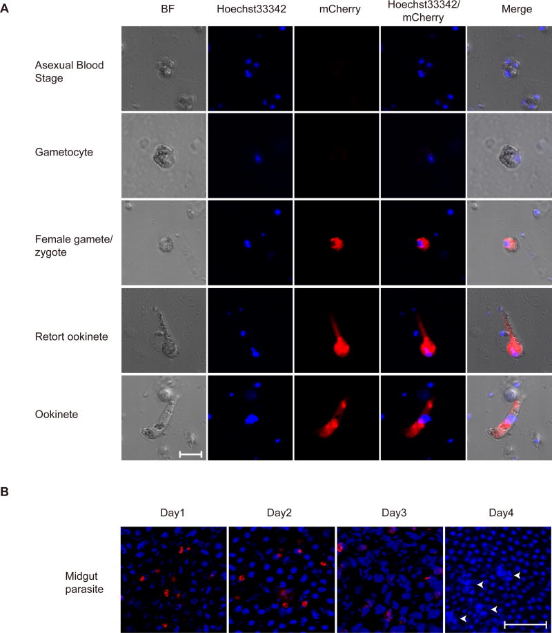
Fig. 3.
Expression of mCherry tagged p28 during different stages of Plasmodium yoelii life cycle. A. Fluorescence microscopy observing of fixed parasites at the indicated stages of life cycle. Nuclei were stained with Hoechst33342 (blue). Results are representative of three independent experiments. Bar=5 µm. B. mCherry-expressing ookinetes and oocysts from infected mosquito midgut. Nuclei were stained with Hoechst33342 (blue). The white arrows mark oocysts in sporogony proliferation with DNA replication. Results are representative of three independent experiments. Bar=5 µm.

