
Fig. 4. Ab initio SAXS reconstruction of Tim50IMS.
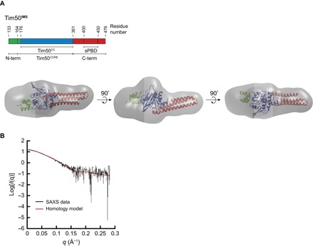
(A) Tim50IMS SAXS molecular envelope aligned with the homology model. The ab initio envelope generated by DAMAVER analysis of SAXS data from Tim50IMS (0.5 mg/ml; gray) is shown in comparison with the Tim50IMS homology model (ribbon structure), aligned using SITUS. The domains of the Tim50IMS homology model are color-coded with respect to the linear domain representation shown above. (B) Analysis of the fit between the envelope and the homology model. The experimental SAXS scattering data for Tim50IMS (0.5 mg/ml; black) and theoretical scattering data for the Tim50IMS homology model (red) were fit using the CRYSOL package, yielding a χ2 value of 1.12.