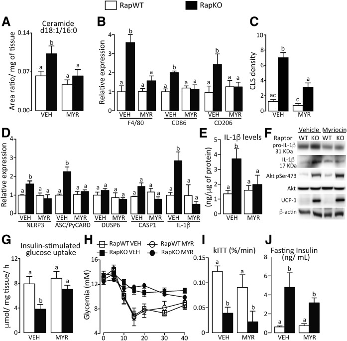
Fig. 5.
MYR abrogated adipose tissue inflammation and inflammasome activation induced by adipocyte raptor deletion. Adipose tissue content of ceramide d18:1/16:0 (A); mRNA levels of macrophage markers (B); CLS density (C); mRNA levels of NLRP3-inflammasome components (D); IL-1β content (E) (ELISA); and pro-IL-1β and mature IL-1β, Akt, and UCP1 contents (F) (Western); adipose tissue insulin-stimulated glucose uptake (G); ITT (H); and rates of glucose disappearance (kITT) (I) and fasting insulinemia (J) in chow-fed RapWT and RapKO mice treated with vehicle (VEH) or MYR (0.5 mg/kg) by gavage for 7 days. Values are mean ± SEM of four to eight mice. Means not sharing a common superscript are significantly different from each other, P < 0.05.

