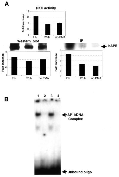
Figure 3.
PMA treatment of APE/Ref-1 stably-transfected K562 cells results in increased amounts of APE/Ref-1 phosphorylation and enhanced redox activity. (A) A monoclonal antibody prepared against APE/Ref-1 was used to immunoprecipitate (IP) the overexpressed protein (and the endogenous expressed APE/Ref-1 as well), and to detect the amount of APE/Ref-1 by western blot as described in Materials and Methods. PKC activity was measured as described in Materials and Methods. (B) The methods for PMA exposure and detection of redox activity by EMSA are provided in the Materials and Methods section. Lane 1, cells exposed to PMA for 2 h; lane 2, immunodepletion of APE/Ref-1 from lysates of cells exposed to PMA for 2 h; lane 3, unexposed cells; and lane 4, immunodepletion of APE/Ref-1 in unexposed cells. The fold increase was quantitated by a densitometer.

