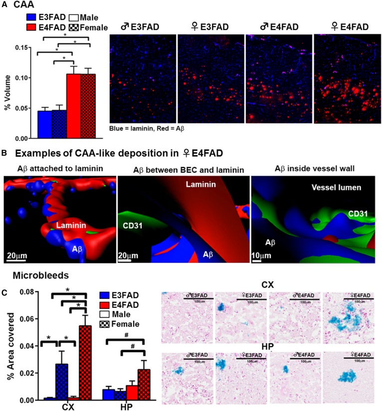
Fig. 7.
CAA and microbleeds in 8-month-old EFAD mice. A: Cortical CAA-like deposition is highest in ♀ E4FAD and ♂ E4FAD; n = 8. Data expressed as mean ± SEM. *P < 0.05 by two-way ANOVA and Tukey’s post hoc comparisons (new data). B: Aβ deposits are found attached to laminin, in the perivascular space and inside the vessel lumen. Examples from ♀ E4FAD mice (new data). C: Microbleeds are highest in ♀ E4FAD mice [adapted from (71)].

