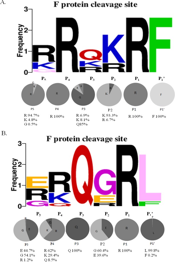
Fig 3. Sequence analysis of Fcs variants at the fusion protein cleavage site in Newcastle disease virus (NDV).
Diversity of residues at each position of the Fcs were analyzed using WebLogo 3.1 (http://weblogo.threeplusone.com/create.cgi). (A) Fcs sequences from 1,073 virulent isolates. (B) Fcs sequences from 499 avirulent isolates. Upper panel: frequency of each residue at a given position. Lower panel: Percentages of each amino acid represented in pie graphs.

