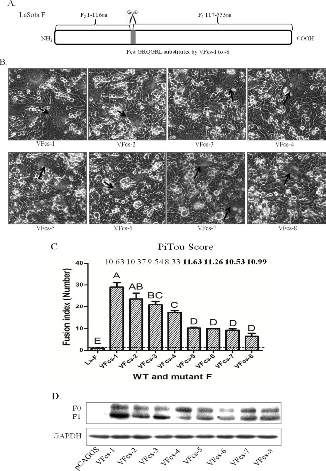
Fig 5. Syncytium formation induced by virulent Fcs and HN in BHK-21 cells.
(A) Schematic of avirulent Fcs substituted with VFcs-1 to VFcs-8 in the LaSota F backbone. (B) Syncytium formation induced by co-transfection with pCAG-Fcs mutants and pCAG-HN of LaSota (0.4 μg of F and 0.4 μg of HN). Syncytia are indicated by black arrows. (C) Quantitation of syncytium formation. BHK-21 cells were co-transfected with pCAG-Fcs mutants and pCAG-HN using Turbofect as described in the Materials and Methods. The number of nuclei in 40 fusion areas was counted at 48 h post-transfection to determine the average syncytia size. Data are the average of syncytia numbers in three independent experiments where statistical significance is designated with capital letters and different letters indicate significant differences (P<0.01). Furin prediction scores of cleavage in Fcs variants were analyzed by Pitou (http://www.nuolan.net/reference.html). (D) Proteolytic cleavage of the F protein with Fcs variants. BHK-21 cells were co-transfected with 0.4 μg of pCAG-F with different Fcs and 0.4 μg of pCAG-HN. The F protein was analyzed via Western blot at 48 h post-transfection. GAPDH protein expression is shown as a control.

