


Fig. 2. Sequence and structure-based alignments for MatA and MatB.
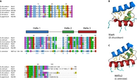
(A) Sequence alignment of MatA and MatB homologs (CLUSTAL color scheme) from a variety of Dictyostelium species shows a high degree of conservation, implying that the structure is very likely to be conserved between these species. The separate lower row shown in the core region is a structure-based alignment of MatA with S. cerevisiae MATα2, which demonstrates that many of the core hydrophobic residues (indicated with blue dots) are also conserved between these two proteins. The structures of MatA (B) and MATα2 (C) show how, in both cases, the side chains of these conserved hydrophobic residues (shown in yellow) are arranged to form the core of the structure. Side chains of solvent-exposed basic residues on the third helix that are likely (MatA) or known (MATα2) to interact with the phosphate backbone of the DNA upon binding are shown in turquoise.