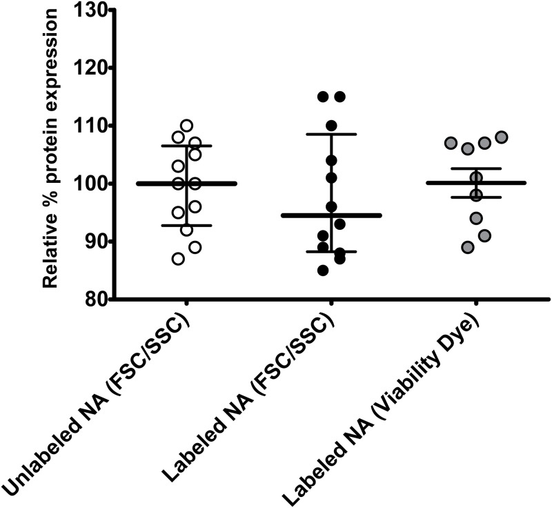
Fig 4. Cellular toxicity can be determined in described flow cytometric method that quantifies transfection efficiency without the use of a viability dye.
293T cells underwent chemical transfection using labeled or standard transfected nucleic acids (pNL4-3 or mCherry plasmids) and the TransITX2 transfection reagent as described in Methods. Gating on viable cells was performed as shown in Fig 1. Each experiment was performed in triplicates and three independent times with specific (un)labelled nucleic acids. Median and interquartile range (IQR) are shown. The non-parametric statistical Kruskal-Wallis test was used for comparisons between groups. Quantification of cell death by forward and side scatter parameters, and doublet discrimination [18.4% (8.6)] gave similar results compared to quantification of cell death by nucleic acid viability dye (7-AAD) [20.50% (11.7)] and amine viability dye [24.4 (11.1)](p = 0.379). Of note use of viability cell dye cannot be used in electroporation experiments (e.g. Jurkat E6 cells electroporation with FITC-labeled DNA mCherry plasmid) due to the mechanisms of action of electroporation methods and dye exclusion tests for cell viability dyes.

