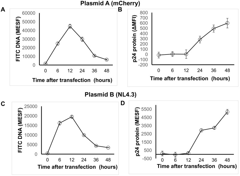
Fig 6. Flow cytometric analysis of DNA uptake and protein expression over time in described flow cytometric method that quantifies transfection efficiency.
293T cells (<20 passages) underwent chemical transfection using the TransITX2 transfection reagent and the mCherry plasmid (plasmid A; A, B) or the NL4.3 plasmid (plasmid B; C, D) as described in Fig 1. Co-expression of DNA taken up by cells (A, C) and target protein (B, D) were analyzed at 6, 12, 24, 36 and 48 hours after transfection. Data are means of triplicates from three independent experiments. Median fluorescence intensity was subtracted from the respective untransfected control (ΔMFI) as described in Methods. Fluorescence intensities were standardized using a MESF standard curve as described in methods. Note that MESF beads are not available for mCherry and in this case the MFI can be used to quantify levels of expression of protein per cell.

