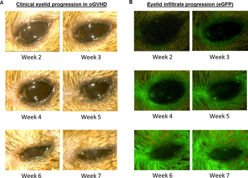
Fig 2. Tempo of clinical and immune infiltrative changes in pre-clinical model of GHVD.

Animals were conditioned with a 10.5 Gy and transplanted with B6-eGFP bone marrow and CD90.1 T-cells. Clinical photographs and eGFP expression was captured by light and in vivo stereo fluorescent microscopy at different time-points post-allogeneic HSCT. A) external photographs demonstrating disease progression at the lid margin. At weeks 4–5, lid edema was present and by week 6, partial lid closure was evident. Severe lid closure occurred by week 7. Clinical examination revealed mild corneal pathology between weeks 6–7 post transplant. B) Fluorescent stereomicroscopy photographs demonstrating increasing eGFP cell infiltrate, with prominent lid margin involvement by week 6. Note the presence of eGFP infiltrate at week 3 correlating with worsening clinical lid edema and closure, with increased infiltrate by weeks 6 and 7. eGFP cell infiltrate localization to the ocular surface precedes development of mild clinical corneal pathology.
