
Figure 2. PDEF regulates cellular proliferation and anti-apoptosis of HCC cell lines.
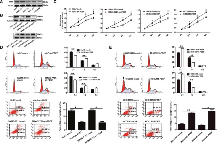
A. PDEF protein is downregulated in HCC cell lines. Western blot was used to quantify the endogenous levers levels of PDEF protein. Tubulin served as the control. B. PDEF expression was confirmed using western blot analysis of stably transfected and of parent cells. C. Cell proliferation tested test by using cell counting kit 8 after knockdown or overexpression of PDEF in HCC cells. Quantitation of was done by reporting the observed OD values for tumor cell OD value. D. Huh7 and SMMC-7721 were transfected with PDEF-shRNA or mock for 48 hours then analyzed by flow cytometry. E. MHCC97H and HCCLM3 were transfected with PDEF forced expression or mock for 48 hours then analyzed by flow cytometry. Percentages (%) of cell subpopulations at different stages of cell cycles and apoptosis are list within the figures. Data was reported as the mean+ values with standard deviations from least three independent experiments, with each assay was performed in triplicate times. *P<0.05, **P<0.01.