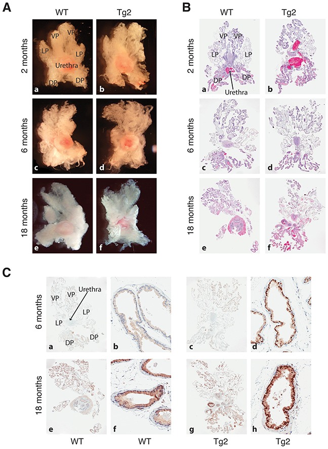
Figure 2. Prostate morphologies in WT and Tg2 mice at different ages.

(A) Representative images of microdissected prostates in WT and transgenic mice at 3 different ages. (B) Representative H&E images of whole-mount prostate sections of WT and Tg2 mice at 3 ages. (C) Representative IHC images of NanogP8 staining in whole-mount prostate sections in WT and transgenic mice at 6 and 18 months of age. Shown on the left (panels a, c, e, g) are low-mag images (40×) and on the right ventral lobes at high magnifications (200×; panels b, d, f, h).
