
Figure 1. Survival fraction, proliferation and DNA damage of lung cells and their daughter cells (DCs).
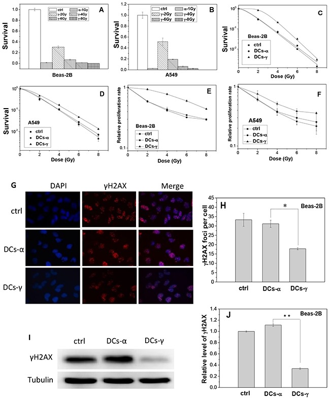
Beas-2B cells and A549 cells were irradiated with priming doses of γ-rays and α-particles, respectively. Then the irradiated cells were cultured for 2 weeks to obtain their DCs. (A, B) Clonogenic survivals of Beas-2B and A549 cells irradiated by 1 Gy α-particles or 2, 4, 6, and 8 Gy γ-rays. (C, D) Clonogenic survivals of DCs-α, DCs-γ and its parent control irradiated with different test doses of γ-rays. (E, F) Proliferation rate of DCs-α, DCs-γ and its parent control irradiated with different test doses of γ-rays. (G, H) γH2AX foci in the DCs-α and DCs-γ of Beas-2B cells. The DCs were irradiated with a test dose of 2 Gy γ-rays and fixed immediately after irradiation for immunostaining assay of γH2AX foci. The foci were counted in at least 200 cells. (I, J) Protein expressions of γH2AX in the DCs-α, DCs-γ and its parent control. Proteins were determined by Western blotting and normalized to its corresponding level of β-Tubulin. Data were presented as means ± SEMs of three independent experiments. * P < 0.05.