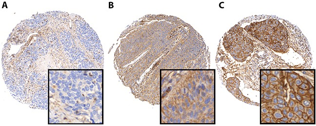
Figure 1. PD-L1 immunohistochemistry in HNSCC.

Representative images of HNSCC demonstrating negative (A), low (B), and high (C) PD-L1 protein levels.

Representative images of HNSCC demonstrating negative (A), low (B), and high (C) PD-L1 protein levels.