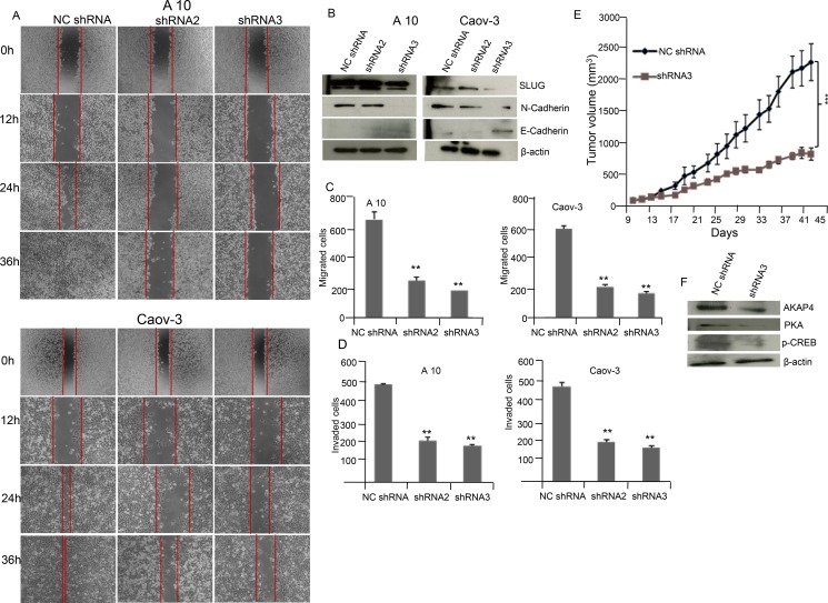
Figure 5. AKAP4 knockdown inhibits wound healing ability and cellular motility marker in A10 and Caov-3 cells and reduces tumor growth in SCID mice model.
(A) Phase contrast microscopy shows decreased wound healing ability in A10 and Caov-3 cells after NC shRNA, shRNA2 and shRNA3 treatment. (B) Western blot shows decreased expression of mesenchymal marker and increased expression of epithelial marker in A10 and Caov-3, β- actin serves as loading control. (C and D) Bar diagram shows significant reduction in migrated and invaded cells after AKAP4 ablation in A10 and Caov-3 cells compared to NC shRNA treated cells. (E) Tumor volume was reduced after shRNA3 treatment significantly (P = 0.001) in SCID mice model compared to NC shRNA treated mice. (F) Western blot shows decreased expression of AKAP4, PKA and p-CREB in shRNA3 treated tumor lyaste. β- actin serves as loading control. The data shown as mean ± standard error of the mean (SEM) of two independent experiments. *P < 0.05; * * P < 0.01, * * * P < 0.001.

