
Figure 4. The role of SREBP1 in p21 expression and cell growth in PDA cells.
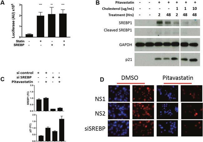
(A) PANC1 cells were transfected with an SREBP1 reporter containing multimerized SRE sites upstream of luciferase. Luciferase activity was measured 48 hours after treatment +/− pitavastatin and +/− a plasmid overexpressing SREBP1a. (B) Cells were treated with10μM pitavastatin for 48 hours and with 0, 1 or 10 μg/mL cholesterol for 2 or 48 hours. Lysates were collected for western blots for: SREBP1- full length and cleaved active form, p21 and GAPDH. (C) Cells were transfected with non-specific (NS) or SREBP1 specific siRNA in the presence or absence of pitavastatin (pita) and harvested for RNA extraction 72 hours later. (D) Concurrently cells were fixed and immunostained for Ki67 (red) and nuclei were stained with DAP1 (blue). Images are 200x. Data are represented as mean +/−SD.