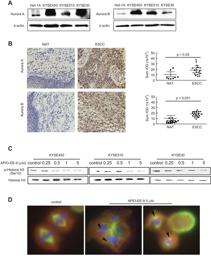
Figure 3. Esophageal cancer cells and tissues highly express Aurora A or Aurora B kinase.

(A) KYSE450, KYSE510 or KYSE30 esophageal cancer cells and normal Het-1A esophageal cells were harvested and Aurora A and B expression was detected by Western blot. (B) Immunohistochemical (IHC) staining of Aurora A and B in esophageal tumor tissues. The integrated optical density (IOD) was evaluated using the Image-Pro Plus software (v. 6.2) program. The asterisks (*, **) indicate a significant difference (p < 0.05, p < 0.01) in tumor staining of Aurora A or B compared with the adjacent normal esophageal tissue (NAT). (C) KYSE450, KYSE510, or KYSE30 esophageal cancer cells were treated with different concentrations of APIO-EE-9 for 24 h. Cells were harvested and phosphorylation of histone H3, a downstream substrate of Aurora B, was detected by Western blotting. (D) APIO-EE-09 induced polyploidy and multiple centrosome formation in esophageal cancer cell lines. KYSE450 cells were stained with DAPI, γ-tubulin, or α-tubulin, after treatment with 5 μM APIO-EE-9 for 2 h (nucleus = blue; centrosome = green; cytoskeleton = red).
