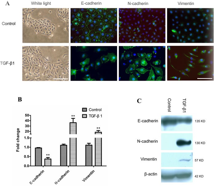
Figure 1. EMT of human immortalized bronchial epithelial cells induced by TGF-β1.

M-BE cells were treated with human recombinant TGF-β1 at a final concentration of 5 ng/ml for 6 days, with cells cultured without TGF-β1 as control. A. A phenotypic change in M-BE from epithelial to spindle-shaped morphology was observed after TGF-β1 treatment, which was photographed at 100× magnification using white light microscopy (left panel, scale bar = 100 μm). Immunofluorescence staining showed the expression status of three EMT markers (left to right panels: E-cadherin, N-cadherin, Vimentin) in M-BE cells induced by TGF-β1. FITC (green) was used for respective target proteins; 4,6-diamidino-2-phenylindole (DAPI) was used to visualize nuclei. All of the fluorescence images were captured at 400× magnification using fluorescence microscopy (scale bar = 25 μm). B. qRT-PCR analysis for mRNA levels of three EMT markers in TGF-β1 treated cells. Y-axis indicates the relative expression level (Fold Change, FC) of genes. Means and standard deviations (SD, error bars) are shown. Unpaired Student's t-test (two sided) was performed for significance estimate. **M-BE cells treated with TGF-β1vs control, P < 0.05. C. Western blotting shows the protein expression levels of E-cadherin, N-cadherin and Vimentin. β-actin is presented for equivalent loading control.
