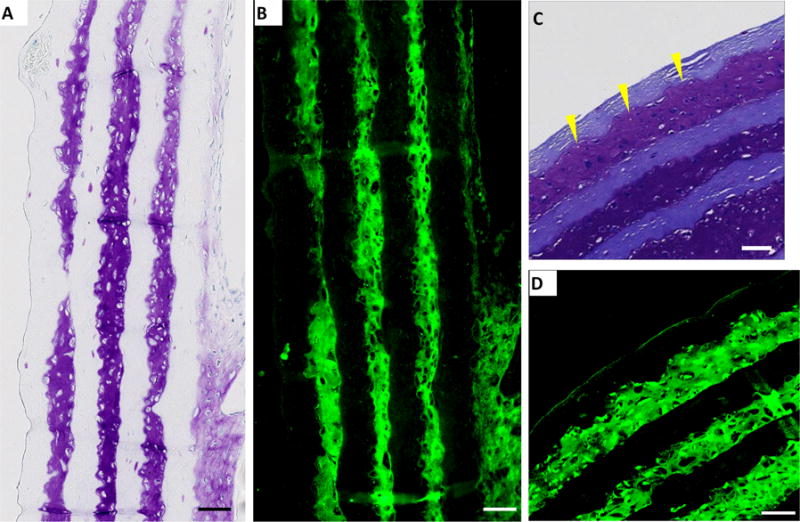
Figure 3.

Immunohistochemistry results. Sample constructs were sectioned in (A, B) longitudinal and (C, D) cross-sectional and stained for collagen type II (green, B, D) using immunochemical methods. Toluidine blue stained sections (A, C) are shown for comparison. A and B are sections from control constructs with no microchannels, C and D are sections from constructs containing 100 micrometer-wide channels with 100 micrometers spacing. Scale bar: 100 micrometers.
