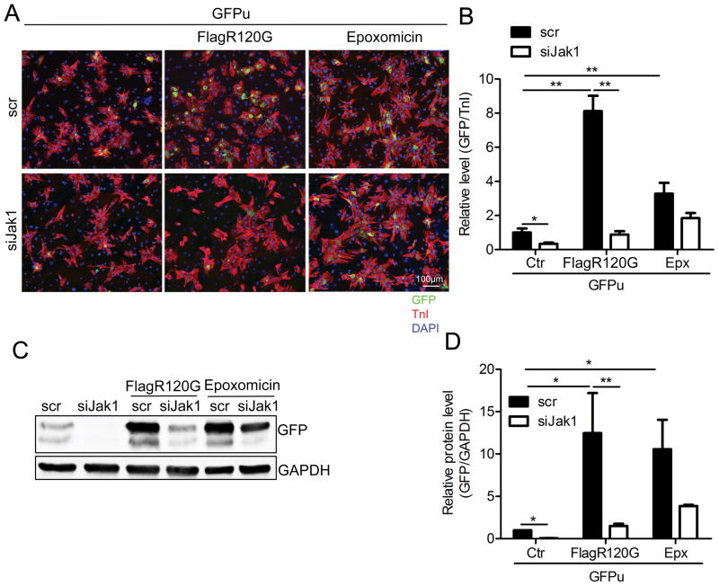
Figure 6. Proteasomal activity is enhanced after Jak1 knockdown.
NRVMs were co-infected with adenoviruses containing Flag-CryABR120G and GFPu (a proteasome activity reporter). The cells were then transfected with siJak1 or a scrambled siRNA (scr) for 4 days and treated with the proteasome inhibitor epoxomicin for 15 hours. At 5 days post-infection, the cells were collected. A, Immunofluorescence with the cardiomyocyte marker TnI (red). Nuclei were counterstained with DAPI (blue). B, The relative GFP level in cardiomyocytes was quantitated using NIS-elements software. Control cells (ctr) were transfected with GFPu adenovirus only. *P<0.05, **P<0.01. C, Western blot analysis of GFP expression level. GAPDH was used as a loading control. D, The Western blot was quantitated. *P<0.05, **P<0.01.

