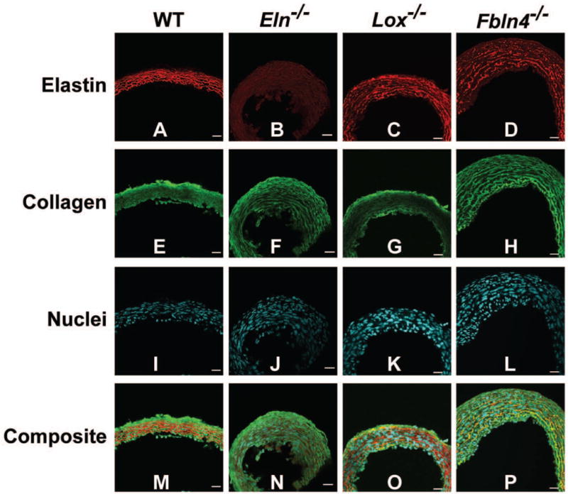
Fig. 4.

Cross-sectional images of newborn AAs. Elastin is red, collagen is green, and cell nuclei are cyan. Elastic lamellae are uninterrupted in WT AA, absent in Eln−/− AA, and fragmented in Lox−/− and Fbln4−/− AA (A – D). Collagen appears normal in KO AAs (E – H). The cell density is similar across groups, but the nuclei appear disorganized near the lumen of KO AAs (I – L). Composite images are shown in panels M – P. The lumen is at the bottom of the images. Scale bar = 20 μm. 3 – 5 aortic sections were examined for each group.
