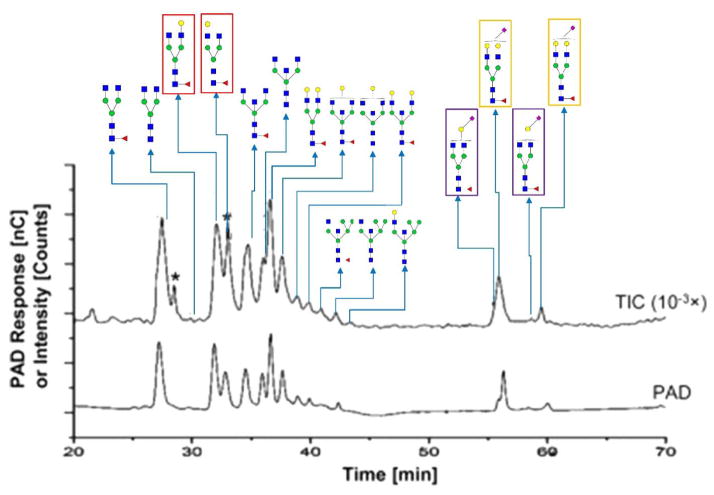
Figure 5.
Isomeric separation performed by HPAEC. HPAEC-PAD chromatograms and total ion current chromatograms from positive-mode HPAEC-MS (intensity attenuation: 10−3×) demonstrating the isomeric separation of PNGase F-released N-glycans from a purified human IgG (h-IgG, 99%). Six branch isomers are highlighted by frames with different colors. The same frame color denotes the isomers from the same glycan composition. Symbols in accordance with CFG notation. A spike peak is indicated by an asterisk. Reprinted and modified from [33].

