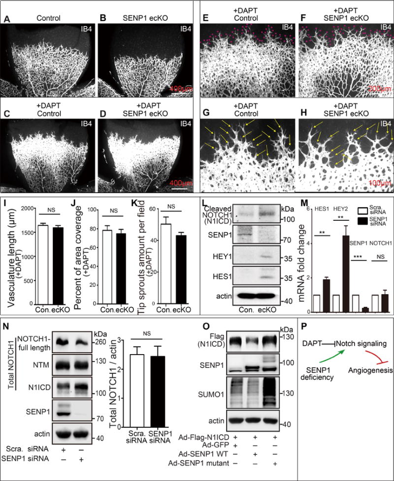
Figure 2. Loss of SENP1 function enhances endothelial NOTCH1 responses.

(A–K) NOTCH response inhibitor, DAPT, completely rescues defects in the retinal vasculature of SENP1-ecKO pups. (A, B) Isolectin B4 (IB4) stained vascular plexus in P6 retina whole mounts from control or SENP1-ecKO mice without DAPT treatment (n=7 and n=8 respectively) or (C–H) with daily injection of DAPT (100 mg kg−1) from the P4 stage (n=9 and n=7 respectively). Red asterisks indicate the tip sprouts of vessels and yellow arrows indicate tip cells. (I–K) Statistical summary of vascular parameters in retinas from each genotype. (L–O) SENP1 limits endothelial NOTCH signaling in vitro and in vivo. (L) Protein expression of NOTCH signaling in MLEC isolated from control or SENP1-ecKO mice, and (M) endothelial NOTCH signaling was determined by measuring the mRNA levels of NOTCH1 intracellular domain (N1ICD) and NOTCH1 target genes (HES1 and HEY2), and SENP1 in control (scramble) or SENP1-siRNA-transfected HMVEC cells. (M–N) SENP1 deficiency does not affect endothelial NOTCH1 expression on mRNA (M) and protein levels (N). (O) N1ICD stability was tested in HUVEC bearing SENP1 wild-type or catalytic inactive mutant. (P) A model for a critical role of SENP1 in regulation of endothelial NOTCH1 signaling and NOTCH1-dependent angiogenesis.
