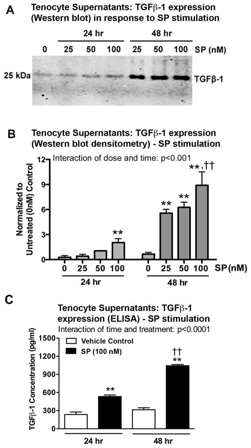
Figure 3.
SP increases TGFβ-1 expression in supernatants of cultured rat tenocytes in dose and time dependent manners. Equal numbers of passage 3–5 tenocytes were plated and grown to 90% confluence, serum restricted (1–2% FBS) for 24 hours, and treated with SP at indicated concentrations for 24 or 48 hours. (A) Representative Western blot of tenocyte supernatants probed with anti-TGFβ-1. (B) Densitometric results of Western blots from four independent experiments ± SEM. TGFβ-1 band was normalized to time-matched vehicle control levels (PBS and 1% BSA only; indicated as “0”). (C) TGFβ-1 expression in tenocyte supernatants was assessed using ELISA at 24 and 48 hours after onset of SP treatment (using the 100 nM dose). Data are the means of eight independent experiments ± SEM. ANOVA results shown in panels B–D. **: p<0.01, compared to time-matched vehicle controls. ††: p<0.01, compared to 24-hour time-point.

