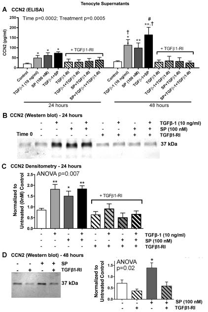
Figure 5.
SP induces CCN2 expression via the TGFβ-1 signaling pathway in rat tenocyte supernatants. Equal numbers of passage 3–5 tenocytes were plated and grown to 90% confluence, serum restricted (1–2% FBS) for 24 hours, and then treated with TGFβ-1 (10 ng/ml), SP (100 nM), or DMSO (vehicle for TGFβ1-RI, indicated as Control or -) in the absence or presence of TGFβ1-RI (500 nM) for 24 or 48 hours. (A) CCN2 production assessed using ELISA. Data are means of seven independent experiments ± SEM. (B) Representative Western blot of tenocyte supernatants probed with anti-CCN2 after stimulating for 24 hours. (C) Densitometric results of five independent experiments ± SEM, after stimulating for 24 hours. CCN2 band was normalized to 24 hour vehicle control levels (DMSO). (D) Left: Representative Western blot of tenocyte supernatants probed with anti-CCN2 after stimulating for 48 hours. Right: Densitometric results of three independent experiments ± SEM. CCN2 band was normalized to 48 hour vehicle control levels (DMSO). ANOVA results shown in panels A, C and D. * and **: p<0.01, respectively, compared to vehicle control levels; #: p<0.05, compared to SP treatment; †: p<0.05, compared to 24 hour time-point.

