
Figure 2.
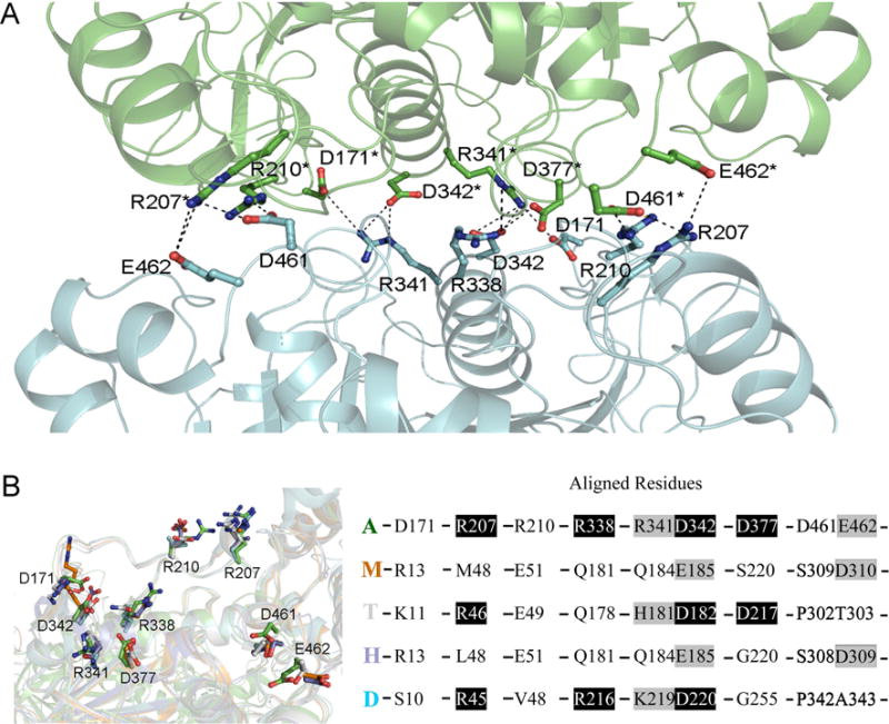
(A) Salt bridges in the intermolecular interface of dimer 1 in structure are shown with dashed lines, the involved residues are shown in sticks, and other part of the dimer is shown in cartoon presentation. (B) Superposition of AgAChE chain A (A, green), hAChE (H, 4ey4, blue), mouse AChE (M, 2ha2, brown), TcAChE (T, 2wg2, grey), and Drosophila AChE (D, 1qo9, cyan). The left panel shows the residues (in sticks) of the four other structures structurally aligned with the residues involved in salt bridge formation in AgAChE CD dimer 1; the right panel shows the residues in AgAChE CD involved in salt bridge formation and the aligned residues from other AChEs (black background: same residues, grey background: similar residues).