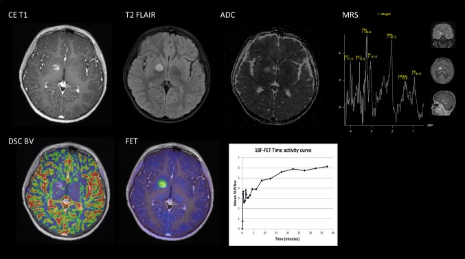
Fig. 1.
Multimodality tumor characterization. A combined 40 min dynamic FET PET/MRI with DSC BV and single voxel MRS was performed in a 7-year-old boy with an incidentally found lesion in the right basal ganglia area. Post-contrast T1 (CE T1) and T2 FLAIR show a solitary contrast-enhancing lesion without edema. Supplementary imaging included: DWI showing high ADC and thus not indicative of increased cellularity; MRS (short echo time) demonstrating only moderately increased choline (Cho/NAA = 1.36 and Cho/Cr = 1.23); leakage-corrected DSC BV did not show increased BV; dynamic FET PET scanning found moderately increased uptake (T max/B = 2.1) with an increasing time–activity curve. Based on the combined imaging, a differentiation of a neoplasm from inflammatory (or other non-neoplastic) pathology could not be made, but it was concluded that a high-grade glioma or other aggressive malignancy was unlikely. Follow-up MRI after 3 months showed regression of contrast enhancement pointing toward a demyelinating lesion

