Fig. 4.
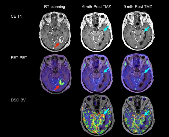
Tumor recurrence vs. treatment effects. Transaxial T1-weighted post-contrast MRI (top row CE T1), FET PET (center row) and leakage-corrected blood volume maps (bottom row DSC BV) in a patient with deep-seated glioblastoma multiforme (WHO IV) in the left inferior occipito-temporal lobe. The initial scans at radiotherapy planning (left) show 6 cm3 of metabolically active tumor with FET uptake (red arrow, T max/B = 2.7). Six months after termination of adjuvant temozolomide (TMZ), CE T1 MRI found increased contrast enhancement in the left hippocampus suspicious of tumor recurrence, and the patient was scheduled for second-line chemotherapy. However, recurrence could not be corroborated by supplementary FET PET/MRI DSC scanning (cyan arrow, T max/B = 1.4). Evidence of tumor angiogenesis could not be identified with certainty because of the high blood volume signal from surrounding vasculature and choroid plexus. Follow-up FET PET/MRI DSC after 3 months untreated showed stable conditions (right column) supporting treatment effects

