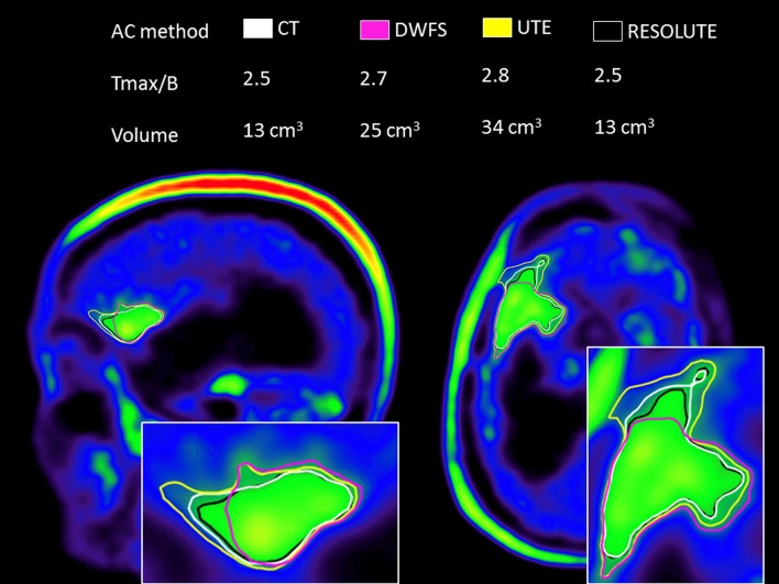
Fig. 6.
PET/MRI attenuation correction. Sagittal and axial image of simultaneous 18F-FET PET/MRI acquisition of a 55-year-old female with anaplastic oligodendroglioma (WHO III) with the tumor borders delineated by activity >1.6 times the background in the healthy brain. The PET reconstruction is performed applying four different attenuation correction strategies using either CT (a low-dose CT performed on a separate PET/CT scanner), Dixon water–fat separation (DWFS), ultrashort echo time (UTE) or RESOLUTE that identifies bone signal in the MRI. RESOLUTE (white) most accurately resembles CT attenuation correction (black) regarding both tumor volume and maximal tumor uptake relative to a background region (T max/B). DWFS and UTE significantly overestimate volume and signal intensity and warp the configuration of the tumor due to radial error. The images were kindly provided by Claes Nøhr Ladefoged

