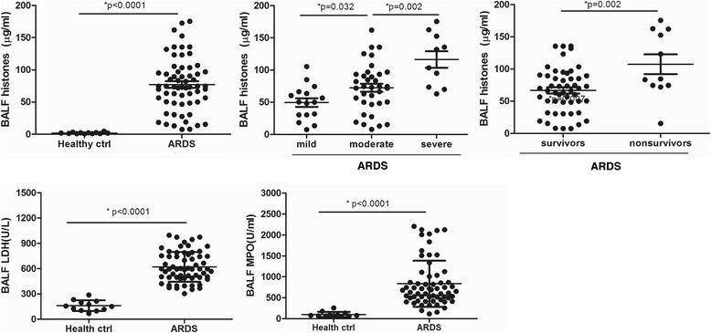
Fig. 2.

Extracellular histones were detected in the BALF of ARDS patients and their possible sources. Extracellular histones were also significantly higher in the BALF of ARDS patients than in healthy controls. Likewise, severe ARDS patients had higher BALF extracellular histone levels than moderate or mild patients, whereas non-survivor ARDS patients had higher BALF extracellular histone levels than survivors. Quantification of BALF LDH activity, a marker reflecting tissue damage, and BALF MPO activity, a marker of inflammatory cell activation, showed that both LDH and MPO levels were increased remarkably in ARDS patients compared to healthy controls, thus indicating a possible cellular sources for extracellular histones. Variables were expressed as median (interquartile range)
