Fig. 6.

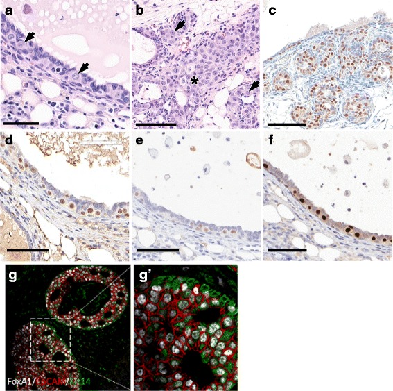
Large oval pale (LOP) cells in 129:Stat1 −/− mammary glands developing cancer. The figure illustrates the potential cancer-initiating LOP cells in 129:Stat1 −/− mammary intraepithelial neoplasia (MIN) and tumors. The LOP cell has a pale cytoplasm and a large oval nucleus with clear chromatin. These cells initially appear in ducts before MIN are detectable. a Hematoxylin and eosin (H&E) staining of duct shows LOP cells standing out from the normal luminal layer (arrows). The normal mammary epithelial cells (MECs) have densely staining nuclei and relatively sparse cytoplasm. These LOP cells were found only in aging 129:Stat1 −/− females with tumors or MIN. Scale bar = 100 μm. b H&E-stained image shows LOP cells filling a duct (asterisk) and populating side buds (arrows). Scale bar = 100 μm. c Progesterone receptor (PR)-stained tissue shows side budding that is forming an early MIN lesion. Scale bar = 70 μm. d Forkhead box A1 (FoxA1)-positive LOP cells were observed in a duct from a tumor-bearing mouse. An adjacent duct was negative for FoxA1. Scale bar = 70 μm. e and f Immunohistochemical (IHC) stains show uniformly positive LOP cells for (e) estrogen receptor (ER) and (f) PR. FoxA1+/ER+/PR+ LOP cells in tumor are shown in Additional file 2: Figure S6. g and g′ A multiplex IHC image shows FoxA1 (white), epithelial cell adhesion molecule (red), and keratin 14 (green) expression in MIN in 129:Stat1 −/−. g′ Higher-magnification image of inset shown in g. Note the dual staining for basal (green) and luminal (red) antigens in many of the LOP cells, indicating that they are dual-staining and potentially pluripotential
