Fig. 10.

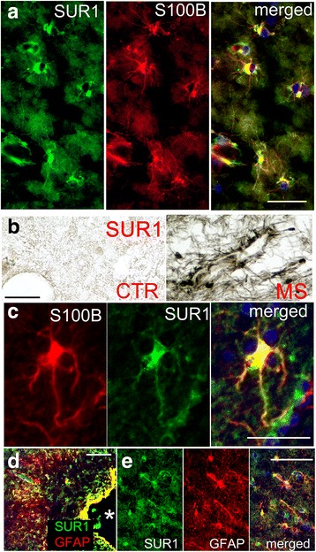
SUR1 is upregulated in astrocytes in demyelinating MS lesions. a Co-immunolabeling for SUR1 (green) and S100B (red) in a chronic active lesion; merged images are indicated; representative of 13 active, chronic active, or chronic inactive WM lesions. b, c Perivascular astrocytes, shown at low (b, right) and high (c) magnification, extend SUR1-positive processes toward nearby vessels, as visualized by chromagen (b) and fluorescent (c) immunolabeling; note absent SUR1 expression in control white matter (b, left). d, e Co-immunolabeling for GFAP (red) (d) and SUR1 (green) in a subpial/cortical demyelinating lesion, shown at low (d) and high (e) magnification; asterisk, bottom of a sulcus; representative of 6 cortical lesions; scale bars 50 μm (a, c) and 100 μm (b, d)
