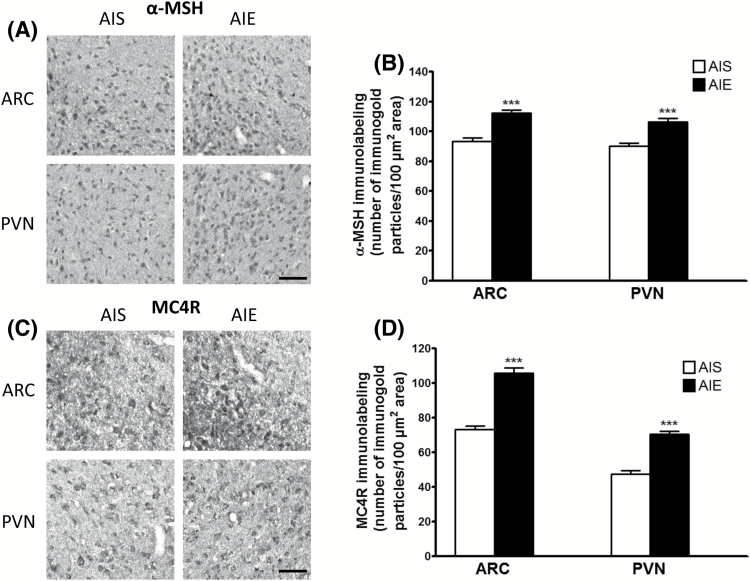
Figure 4.
Effects of adolescent alcohol exposure on the protein levels of α-melanocyte stimulating hormone (α-MSH) and melanocortin 4 receptor (MC4R) in the hypothalamus of rats in adulthood. (A) Representative photomicrographs (scale bar = 50 μm) of α-MSH staining in the arcuate nucleus of the hypothalamus (ARC) and the paraventricular nucleus of the hypothalamus (PVN) of adolescent intermittent ethanol (AIE)- and saline (AIS)-exposed adult rats. (B) Bar diagram showing protein levels of α-MSH in ARC and PVN in AIE- and AIS-exposed adult rats as measured by gold immunolabeling. (C) Representative photomicrographs (scale bar = 50 μm) of MC4R staining in the ARC and PVN in AIE- and AIS-exposed adult rats. (D) Bar diagram showing protein levels of MC4R in ARC and PVN of AIS and AIE adult rats as measured by gold immunolabeling. Values are represented as the mean (±SEM) of the number of immunogold particles/100 μm2. Values are significantly different from AIS-exposed adult group (***P<.001, Student’s t test, n = 6).

