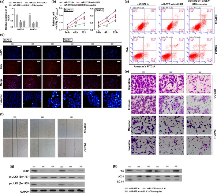
Figure 6.

Autophagy inhibitor and ULK1 knockdown reversed the effects of miR‐372 inhibition in HPAC cells. BXPC‐3 and PANC‐1 cells were transfected with miR‐372 inhibitor, co‐transfected with ULK1 siRNA and miR‐372 inhibitor, or co‐transfected with ULK1 siRNA and miR‐372 inhibitor and additionally stimulated with chloroquine. (a) ULK1 expression on mRNA levels by qRT‐PCR. (b) Cell proliferation rates were determined by the CCK8 assay. (c) Cell apoptosis was determined flow cytometry. (d) Cell proliferation and apoptosis were detected by EdU (Scale bar: 100 μm) and Hoechst 33 258 (400 × magnification) staining assay. (e,f) Migration and invasion were determined by transwell migration (200 × magnification) assay, transwell invasion assay (200 × magnification) and wound scratch assay (40 × magnification). (g) Total protein and phosphorylation levels of ULK1 were determined by western blotting. (h) P62 and LC3‐II level were determined by western blotting. GAPDH was used as an internal control. *P < 0.01 versus miR‐372 inhibitor group.
