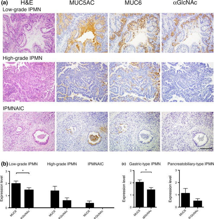
Figure 2.

Immunohistochemical analysis of MUC5AC, MUC6 and αGlcNAc in IPMN and IPMNAIC. (a) MUC5AC is expressed in tumor cells, irrespective of histological grade. MUC6 is highly expressed in tumor cells showing a pyloric gland phenotype characteristic of low‐grade IPMN. However, MUC6 expression decreases in high‐grade IPMN and IPMNAIC. αGlcNAc expression in low‐grade IPMN coincides with that of MUC6. By contrast, in high‐grade and IPMNAIC, αGlcNAc is not expressed in MUC6‐positve tumor cells. Bar = 100 μm. (b) Semi‐quantitation of MUC6 and αGlcNAc expression in low‐grade IPMN, high‐grade IPMN and IPMNAIC. Data are represented as the mean ± SEM. *P < 0.05 by Wilcoxon matched‐pair test. (c) Semi‐quantitation of MUC6 and αGlcNAc expression in gastric‐type IPMN and pancreatobiliary‐type IPMN. Data are represented as the mean ± SEM. *P < 0.05 by Wilcoxon matched‐pair test.
