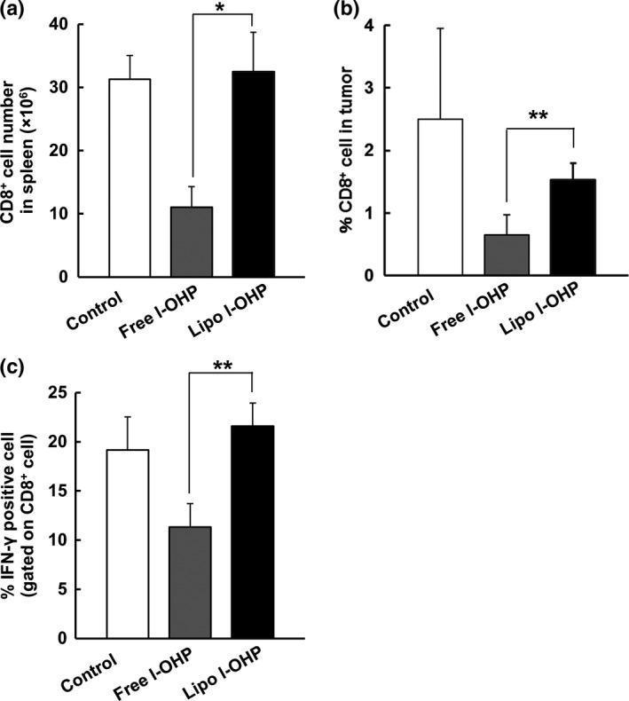
Figure 4.

Liposomal l‐OHP CD8+ T cell‐mediated antitumor immunity. On days 0, 7 and 14, C26 tumor‐bearing BALB/c mice received three intravenous injections with either Free l‐OHP or Liposomal l‐OHP. Non‐treated mice served as the control. On day 21, the tumors and spleens were collected, and cell suspensions were prepared. The suspensions were stained with anti‐CD8 antibody and then analyzed using flow cytometry. (a) The number of CD8+ T cells in the spleens. (b) The frequency of CD8+ T cells in tumors. The percentage of CD8 + T cells was calculated by dividing CD8 + T cell number by total cell number in tumor tissue. (c) On days 0 and 7, C26‐bearing BALB/c mice received two intravenous injections of either Free l‐OHP or Liposomal l‐OHP. Non‐treated mice served as the control. On day 10, the spleens were collected and cell suspensions were prepared. The cells were pulsed with mitomycin C‐treated C26 tumor cells in vitro. IFNγ+ CD8+ T cells were analyzed through flow cytometry. Each value represents the mean ± SD (n = 3). *P < 0.05, **P < 0.01.
